
Ultrasound-guided filler injection: what patients should know
Discover how ultrasound technology is enhancing safety in aesthetic medicine by allowing practitioners to visualize facial anatomy during dermal filler procedures.

Hinweis zu KI-generierten Inhalten (EU-KI-Verordnung Art. 50). Die Autorin oder der Autor hat erklärt, dass dieser Artikel von künstlicher Intelligenz erstellt oder wesentlich unterstützt wurde. Er wurde vor der Veröffentlichung von unserer Redaktion geprüft; die inhaltliche Verantwortung für Richtigkeit und klinische Validität liegt weiterhin bei der Autorin oder dem Autor.
Ultrasound imaging
Ultrasound imaging is transforming the landscape of aesthetic medicine by providing practitioners with a real-time view beneath the skin. This guide explains how this technology supports safer outcomes for those considering or receiving dermal filler treatments. Scan Your Filler is an information platform and does not give medical advice.
Ultrasound-guided injections
Ultrasound-guided injection involves using a high-frequency probe to visualize the internal structures of the face before and during a treatment. By displaying soft tissue and blood vessels on a monitor, the practitioner gains a clearer understanding of the terrain under the skin. This visual feedback assists the practitioner in making informed decisions about the placement of products.
Pre-treatment mapping
Before any filler is injected, a practitioner may use ultrasound to map the specific anatomy of the area. This process helps identify the precise location of sensitive structures such as nerves and arteries, which can vary significantly between individuals. Mapping allows the practitioner to plan a pathway that minimizes the risk of accidental vessel puncture.
Existing filler
Many patients have undergone previous aesthetic treatments that may have left residual filler within the tissues. Ultrasound is a valuable tool for identifying the location, volume, and condition of this older product. Knowing exactly where previous material remains helps the practitioner avoid complications and ensure that new treatments are placed appropriately.
Individual anatomy
Every face possesses unique anatomical variations that are not always visible on the surface. Ultrasound allows the practitioner to see these internal differences, such as the specific depth and path of vital blood vessels. This level of insight enables a highly personalized approach, as the practitioner can adjust their technique to suit the patient's actual internal structure.
Appointment expectations
During an ultrasound-assisted session, the practitioner will apply a water-based gel to the skin and glide a probe over the treatment area. You will likely see the scan displayed on a screen as the clinician evaluates the anatomy and verifies the position of the filler. This process typically adds a short period to the consultation time but provides significant clarity regarding the procedure.
Qualified practitioner
When choosing a professional, look for evidence of specialized training in musculoskeletal or aesthetic ultrasound. A qualified practitioner should be able to explain why they are using the scan and how it benefits your specific treatment plan. Always verify that your provider is a registered medical professional with a clear understanding of facial anatomy.
If you are interested in discussing ultrasound-assisted treatments, you can find trained professionals in our global directory of ultrasound-trained practitioners.
Is an ultrasound scan always required for a filler procedure?
While ultrasound is a powerful tool for safety, it is not required for every patient or every type of filler treatment. The decision to use imaging is always based on the individual clinical judgment of your practitioner.
Does an ultrasound scan hurt?
No, an ultrasound scan is a non-invasive procedure that involves a probe gliding over the skin with gel. It is completely painless and does not involve radiation or needles.
Can ultrasound help if I am worried about complications?
Ultrasound is highly effective for monitoring the integrity of filler and detecting potential issues early. If you have concerns about previous injections, a scan can provide a clear view of the area and help your practitioner determine the best course of action.
What should I ask my practitioner about ultrasound?
You should ask if they are trained in using ultrasound for aesthetic procedures and how they integrate it into their safety protocols. It is also helpful to ask how the imaging specifically helps them manage your unique facial anatomy.
Weiterlesen
Alle Artikel
 Patient guide
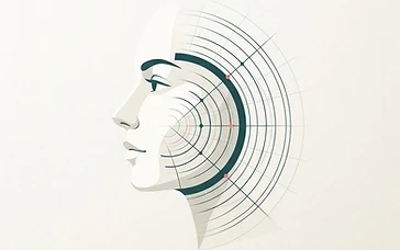
Is my filler still there? What an ultrasound scan shows
Wondering if your dermal filler is still present or has migrated? Ultrasound scans offer a clear, non-invasive way to visualize your fillers and make informed decisions about your care.

 Patient guide
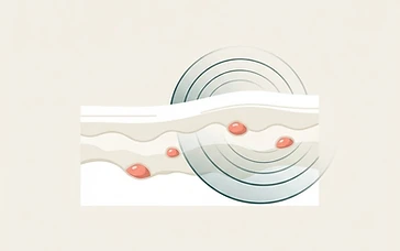
Filler lumps and nodules: what an ultrasound scan reveals
Discover how ultrasound technology helps patients and practitioners understand dermal filler lumps and nodules for informed decision-making.

 Clinical Insights
Why I Never Touch a Syringe Without My Ultrasound Machine
After every severe filler complication I treat with ultrasound guidance, patients ask the same question: 'Why don't other doctors do it this way?' This article is my attempt to answer it.