
Filler lumps and nodules: what an ultrasound scan reveals
Discover how ultrasound technology helps patients and practitioners understand dermal filler lumps and nodules for informed decision-making.
Hinweis zu KI-generierten Inhalten (EU-KI-Verordnung Art. 50). Die Autorin oder der Autor hat erklärt, dass dieser Artikel von künstlicher Intelligenz erstellt oder wesentlich unterstützt wurde. Er wurde vor der Veröffentlichung von unserer Redaktion geprüft; die inhaltliche Verantwortung für Richtigkeit und klinische Validität liegt weiterhin bei der Autorin oder dem Autor.
Finding an unexpected lump or nodule after a dermal filler treatment can be a source of anxiety for many patients. Understanding what these changes represent is the first step toward finding clarity and appropriate professional guidance. Please note that Scan Your Filler is an information platform and does not give medical advice.
Understanding lumps and nodules
Lumps and nodules refer to palpable changes under the skin following a filler procedure. While many of these are temporary and settle on their own, others may persist or appear weeks or months later. They are essentially localized areas where the filler material or the tissue response becomes noticeable to the touch.
Potential causes for filler changes
Several factors can contribute to the formation of these bumps. These include the specific characteristics of the product used, the injection technique applied, or the total volume of material placed in a specific area. Additionally, delayed inflammatory responses or the natural aging process can sometimes lead to visible changes in how filler behaves over time.
The role of ultrasound imaging
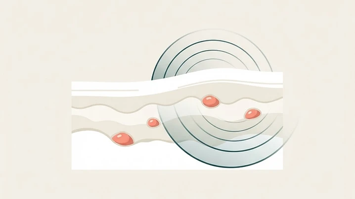
Ultrasound serves as a non-invasive tool that allows practitioners to visualize the layers beneath the skin. It helps differentiate between the filler material itself, inflammation, or other anatomical structures that might be mistaken for a nodule. This imaging provides a map of the area that is invisible to the naked eye.
The scan appointment experience
During a scan, a practitioner applies a conductive gel to the area and moves a small probe over the skin. The process is generally comfortable and provides real-time images on a monitor. The goal is to observe the placement, shape, and surrounding tissue health to gain a better understanding of the situation.
Diagnostic importance before treatment
Using ultrasound before making any decisions about treatment is a crucial step for many practitioners. It provides objective information that guides the conversation between the patient and the provider. By identifying the exact nature of the concern, both parties can discuss potential options with a much clearer understanding of the underlying anatomy.
Questions to ask your practitioner
When discussing your concerns with a professional, consider asking the following questions to help guide the conversation:
- Can you explain what the ultrasound findings represent?
- How does the appearance of this nodule compare to typical filler behavior?
- What are the risks and benefits of different management approaches for this specific case?
- Do we need further diagnostics before considering any changes to the current treatment plan?
Next steps
To locate a professional who utilizes this technology for assessment, you can search our directory of aesthetic ultrasound practitioners.
Is ultrasound always required for filler lumps?
No, ultrasound is a diagnostic tool that a practitioner may choose to use depending on the specific clinical situation. Whether a scan is necessary is a decision that should always be made by a qualified healthcare professional.
Can ultrasound tell if a lump is harmful?
Ultrasound helps the practitioner visualize the structure and location of a lump, which assists them in assessing its nature. It provides essential information that informs their clinical judgment regarding the best path forward.
Does a scan hurt?
An ultrasound scan is typically a comfortable, non-invasive procedure that does not require needles or incisions. Most patients find the experience very straightforward.
Will the scan guarantee a solution?
No, a scan is an imaging tool designed to provide information rather than provide a treatment result. The findings from the scan serve to guide the clinical decision-making process between you and your practitioner.
Weiterlesen
Alle Artikel
 Patient guide
Is my filler still there? What an ultrasound scan shows
Wondering if your dermal filler is still present or has migrated? Ultrasound scans offer a clear, non-invasive way to visualize your fillers and make informed decisions about your care.

 Patient guide
Ultrasound-guided filler injection: what patients should know
Discover how ultrasound technology is enhancing safety in aesthetic medicine by allowing practitioners to visualize facial anatomy during dermal filler procedures.

 Clinical Insights
Why I Never Touch a Syringe Without My Ultrasound Machine
After every severe filler complication I treat with ultrasound guidance, patients ask the same question: 'Why don't other doctors do it this way?' This article is my attempt to answer it.