
Is my filler still there? What an ultrasound scan shows
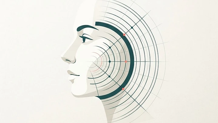
Wondering if your dermal filler is still present or has migrated? Ultrasound scans offer a clear, non-invasive way to visualize your fillers and make informed decisions about your care.
Aviso sobre contenido generado por IA (Reglamento de IA de la UE, art. 50). El autor ha declarado que este artículo fue generado o asistido sustancialmente por inteligencia artificial. Ha sido revisado por nuestro equipo editorial antes de su publicación y el autor mantiene la plena responsabilidad editorial sobre su exactitud y validez clínica.
Ultrasound scan information
Many people find themselves questioning the state of their past dermal filler treatments long after the procedure is finished. An ultrasound scan provides a real-time window beneath the skin to visualize filler deposits and confirm their exact status. Scan Your Filler acts as an independent information platform and does not provide medical advice. Understanding what lies beneath your skin can help you make better informed decisions regarding your aesthetic journey.
Tissue layers and imaging
An ultrasound scan works by using high-frequency sound waves to create detailed images of the various tissue layers. These scans can clearly differentiate between filler deposits, blood vessels, nerves, and surrounding anatomical structures. By seeing exactly where the filler is located, your practitioner can assess its integrity and position without any guesswork.
Dermal filler longevity
Dermal fillers are often designed to resist quick degradation, meaning they can persist in the body for much longer than marketing materials might suggest. Ultrasound imaging often reveals that product remains encapsulated or integrated within the tissue years after it was initially placed. This longevity is common, though it varies significantly depending on the specific type of product used.
Product migration and lumps
When patients notice lumps or unexpected asymmetry, they often worry about product migration. Ultrasound is the gold standard for tracking whether filler has physically moved from its original site or if it has simply shifted due to tissue movement. It can also identify if a lump is composed of filler or if it is a result of something else like scar tissue or a localized reaction.
Scan appointment process
During a scan, a practitioner applies a specialized probe to the target area while viewing images on a screen. The process is non-invasive and generally painless, providing immediate visual feedback about the depth and volume of the material. Your practitioner will explain what they see on the screen to help you understand the current state of your previous treatments.
Dissolving procedures and practitioners
If a scan reveals filler that is contributing to complications, infection, or aesthetic distress, a practitioner might discuss the option of dissolving it. Scanning is essential before any dissolving procedure because it ensures that the product can be accurately targeted and mapped. It prevents the unnecessary use of enzymes on areas where no filler is actually present.
Not all aesthetic professionals have access to or training in ultrasound diagnostics. Look for someone who is experienced in both ultrasound imaging and the management of filler complications. You should feel comfortable asking about their specific training, their experience with ultrasound, and their protocol for dealing with unexpected findings.
Next steps and directory
If you are concerned about your filler, the best course of action is to schedule a consultation with a qualified professional who uses ultrasound technology. You can find experienced practitioners worldwide through our comprehensive directory.
Does ultrasound hurt?
The procedure is non-invasive and typically painless, as it involves only a hand-held probe and a small amount of conductive gel on the skin surface.
Can ultrasound see all types of filler?
Ultrasound can detect most common dermal fillers, although the appearance of different products can vary based on their formulation and density.
Should I get a scan if I have no problems?
While many patients seek scans because of concerns, some individuals choose to get a baseline scan to understand their facial anatomy before pursuing further treatments.
Is dissolving always the right option?
Dissolving is a medical decision that should only be made after a thorough clinical assessment, as it is not always necessary for everyone who has residual filler.
Seguir leyendo
Todos los artículos
 Patient guide
Filler lumps and nodules: what an ultrasound scan reveals
Discover how ultrasound technology helps patients and practitioners understand dermal filler lumps and nodules for informed decision-making.

 Patient guide
Ultrasound-guided filler injection: what patients should know
Discover how ultrasound technology is enhancing safety in aesthetic medicine by allowing practitioners to visualize facial anatomy during dermal filler procedures.

 Clinical Insights
Why I Never Touch a Syringe Without My Ultrasound Machine
After every severe filler complication I treat with ultrasound guidance, patients ask the same question: 'Why don't other doctors do it this way?' This article is my attempt to answer it.