
Ultrasound Scanning Taught Me to Doubt What I Thought I Knew
For the past three years, ultrasound has been part of my daily practice. What changed is not my technique — what changed is what I can see.

For the past three years, ultrasound has been part of my daily practice - not an occasional add-on, but a routine step I now rely on before, during, and after injecting.
Routine practice of ultrasound
What changed is not my technique. What changed is what I can see.
We inject in a plane where vessels, fascial layers, and fat compartments sit within millimetres of one another. And the anatomy we were taught is a model, an average, a useful fiction.
Real patients depart from it constantly: a facial artery running superficially where it "should" be deep, an angular branch crossing where no atlas predicted it, a temporal vessel where I had always assumed safety.
Visible anatomical variability
Ultrasound made that variability visible. It also made something else visible: the near misses.
Every practitioner who adopts imaging goes through the same quiet, uncomfortable phase - scanning a patient and recognising, retrospectively, the injections you performed blind for years, the vessel you passed beside without ever knowing it was there.
Humbling retrospective frights
Those retrospective frights are humbling. They are also the strongest argument for the technique that I know.
I owe this shift entirely to the practitioners who introduced me to it. My gratitude to Leonie Schelke, Peter Velthuis, and Carlos Zito is genuinely without limit.
Teaching the means of evidence
They did not simply teach me to operate a probe. They taught me to doubt what I thought I knew, and gave me the means to replace assumption with evidence - patient by patient, plane by plane.
A different way of practising
That is not a marginal improvement in safety. It is a different way of practising.
About the author
Dr Claude Levy is a qualified oral and maxillofacial surgeon, stomatologist, and aesthetic physician practicing in the Paris metropolitan area.
Seguir leyendo
Todos los artículos
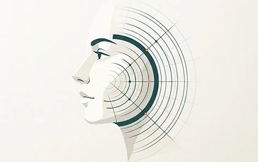 Patient guide
Is my filler still there? What an ultrasound scan shows
Wondering if your dermal filler is still present or has migrated? Ultrasound scans offer a clear, non-invasive way to visualize your fillers and make informed decisions about your care.

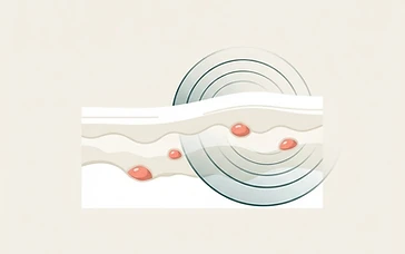 Patient guide
Filler lumps and nodules: what an ultrasound scan reveals
Discover how ultrasound technology helps patients and practitioners understand dermal filler lumps and nodules for informed decision-making.

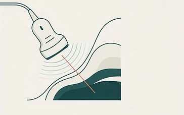 Patient guide
Ultrasound-guided filler injection: what patients should know
Discover how ultrasound technology is enhancing safety in aesthetic medicine by allowing practitioners to visualize facial anatomy during dermal filler procedures.