
Beyond the Surface: A Radiologist's Role in Aesthetic Medicine
Many patients are surprised to learn that what appears completely normal on the surface may tell only part of the story. Important information can remain hidden beneath the skin, even years after an aesthetic treatment.
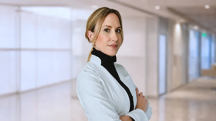
Many patients are surprised to learn that what appears completely normal on the surface may tell only part of the story. Important information can remain hidden beneath the skin, even years after an aesthetic treatment.
Aesthetic imaging practice
As a radiologist specialised in aesthetic imaging, I frequently evaluate patients who are planning a new procedure, have concerns about previous treatments, or require imaging assessment after an unexpected outcome. My work focuses on understanding what lies beneath the surface through high-resolution ultrasound and, when necessary, magnetic resonance imaging (MRI).
Over the years, I have assessed many patients who were unsure about their treatment history. Some could not recall which products had been injected, while others were unaware that filler material was still present years later.
In many cases, imaging revealed findings that could not be detected during a physical examination alone.
High-resolution ultrasound utility
High-resolution ultrasound allows us to visualise fillers, tissue changes, vascular structures, and anatomical variations in real time. This information can be extremely valuable when planning a procedure, particularly in previously treated patients.
Managing aesthetic complications
Imaging also plays an important role when complications arise. Swelling, nodules, asymmetry, delayed reactions, or unexpected treatment outcomes often require an accurate diagnosis before any corrective action can be taken.
In these situations, close collaboration between the treating practitioner and the radiologist is essential.
Multidisciplinary diagnostic approach
One of the aspects I value most about my work is being part of this multidisciplinary approach. By providing a detailed imaging assessment and a formal radiology report, I can help practitioners better understand the underlying anatomy and support safer, more informed clinical decisions.
While ultrasound is often the first-line imaging tool in aesthetic medicine, MRI can provide additional information in selected cases, particularly when evaluating deeper tissues or more complex findings.
Improving treatment decisions
Ultimately, imaging is not only about identifying complications. It is about improving understanding, increasing safety, and helping both patients and practitioners make better-informed decisions.
Because the best decisions are made when we can see what would otherwise remain invisible.
About the author
Dr. Carolina Mariluis — Radiologist specialised in Aesthetic Imaging
Seguir leyendo
Todos los artículos
 Patient guide
Is my filler still there? What an ultrasound scan shows
Wondering if your dermal filler is still present or has migrated? Ultrasound scans offer a clear, non-invasive way to visualize your fillers and make informed decisions about your care.

 Patient guide
Filler lumps and nodules: what an ultrasound scan reveals
Discover how ultrasound technology helps patients and practitioners understand dermal filler lumps and nodules for informed decision-making.

 Patient guide
Ultrasound-guided filler injection: what patients should know
Discover how ultrasound technology is enhancing safety in aesthetic medicine by allowing practitioners to visualize facial anatomy during dermal filler procedures.