
FAQs: Patient Guide to Facial Ultrasound Scanning in Aesthetics
Whether you are considering dermal fillers for the very first time or you have had treatments for years, your safety and the quality of your results are our top priorities. This guide explains how ultrasound improves your care before, during, and after your procedure.

Whether you are considering dermal fillers for the very first time or you have had treatments for years, your safety and the quality of your results are our top priorities. To achieve the best possible outcomes, we use facial ultrasound scanning (USS) in our clinic. This simple, painless technology allows us to look beneath the surface of your skin. This guide will take you through the story of your treatment and explain exactly how ultrasound improves your care before, during, and after your procedure.
Facial ultrasound scanning in aesthetics
Think of an ultrasound scan as a real-time map of your face. Just as an ultrasound is used in medicine to look at organs or monitor a baby, we use a specialised, high-frequency ultrasound device to visualise the layers of your skin, your fat pads, your muscles, and your blood vessels.
Everyone's internal facial anatomy is completely unique. While medical professionals study textbook anatomy, the exact location and depth of your blood vessels can vary wildly from person to person. By scanning your face first, we can see exactly where your major arteries and veins are located. This is called 'vascular mapping' and it allows us to plan a bespoke, safe route for your injections.
The parotid gland can vary in size, and in some people it extends all the way up to the cheeks. Some people also have extra glandular tissue in the mid-face. Filler injected into the parotid gland can lead to inflammation. If you want to understand your individual anatomy, an ultrasound scan provides the right information.
One of the greatest benefits of ultrasound is its ability to locate old filler. Hyaluronic acid fillers hold onto water, making them easily visible on an ultrasound screen. If you have had previous treatments, we can see exactly where that filler is sitting, how much is left, and whether it has migrated from its original position.
This helps us decide if it is safe to add more product or if we need to dissolve the old filler first to give you a natural result. All filler types can be seen on ultrasound, including permanent fillers. People often forget what type of filler was injected in the past. Permanent fillers are best left alone; re-injecting into them can cause problems such as inflammation and abscesses. Ultrasound can locate these fillers so that injections into those areas can be avoided.
The scan is completely painless, non-invasive, safe and uses a simple layer of clear gel on your skin. It only adds a few minutes to your consultation, but it provides vital information to minimise risks and ensure your treatment plan is highly precise.
Guided injections for precision and safety
Historically, aesthetic injections were performed 'blind'. Practitioners relied heavily on their anatomical knowledge and the feel of the tissues. Today, we can use ultrasound to guide the needle or cannula in real time. We can watch the tip of the instrument on the screen and see the filler being safely deposited into the exact right layer of tissue.
The most severe complication in aesthetic medicine is a 'vascular occlusion'. This happens when filler is accidentally injected into a blood vessel or compresses a vessel, blocking the blood supply to the skin. Because we can visualise both your blood vessels and our needle on the screen at the same time, we can navigate safely around your arteries. This dramatically minimises the risk of vascular occlusions, bruising, and swelling.
Guided injections offer unparalleled precision. Because we can see the specific layers of your tissue, we can place the filler exactly where it will provide the best structural support and the most natural lift. This means we often need to use less product to achieve a superior result, preventing the 'overfilled' look.
Managing issues after treatment
Lumps and bumps can occasionally happen after filler treatments, but diagnosing them by touch alone is very difficult. Is it a normal bruise, a harmless cyst, a pocket of swelling, or a nodule of filler? With ultrasound, we do not have to guess. We can scan the area and instantly diagnose the cause of the lump, allowing us to organise the correct treatment immediately.
If a blocked blood vessel is suspected, immediate action is required to restore blood flow. Ultrasound is the gold standard for managing this complication safely. We can use the scanner to locate the exact artery that is blocked and pinpoint the precise droplet of filler causing the issue.
To dissolve hyaluronic acid filler, we use an enzyme called hyaluronidase. Without ultrasound, practitioners have to 'flood' the general area with large amounts of the dissolving enzyme and hope it reaches the blockage. With ultrasound guidance, we can inject a tiny, precise amount of the enzyme directly into the problematic filler deposit. This targeted approach is safer, resolves the issue much faster, and preserves your natural tissue.
Many patients come to us simply for a diagnostic scan. If you feel puffy, asymmetrical, or suspect your old filler has migrated over the years, an ultrasound assessment is the perfect way to evaluate your facial tissues and put your mind at ease.
Your safety and results
By using ultrasound technology, we are replacing guesswork with scientific precision. We believe you deserve the highest standard of medical care in aesthetics, and visualising your unique anatomy is the key to delivering beautiful, safe, and natural results.
For more information about aesthetic ultrasound education & training by Cutaneous, click here.
About the author
This content has been authored by a clinical expert in the field of aesthetic medicine, dedicated to the integration of ultrasound technology to ensure the highest standards of patient safety and precision in every procedure.
Written by
Dr Leonie Schelke MD, Dr Peter Velthuis MD & Mr Vivek Kuvelker MSc
Erasmus Medical Centre, Netherlands & Skinpharma Aesthetics, UK · Netherlands
Keep reading
All articles
 Patient guide
Is my filler still there? What an ultrasound scan shows
Wondering if your dermal filler is still present or has migrated? Ultrasound scans offer a clear, non-invasive way to visualize your fillers and make informed decisions about your care.

 Patient guide
Filler lumps and nodules: what an ultrasound scan reveals
Discover how ultrasound technology helps patients and practitioners understand dermal filler lumps and nodules for informed decision-making.

 Patient guide
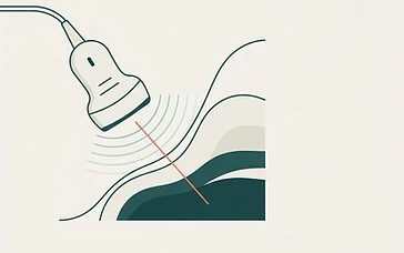
Ultrasound-guided filler injection: what patients should know
Discover how ultrasound technology is enhancing safety in aesthetic medicine by allowing practitioners to visualize facial anatomy during dermal filler procedures.